First, some background
External electrical stimulation of the brain may improve cognitive, motor and memory performance, assist stroke rehabilitation and treat epilepsy. Ideally, electrical stimulation should guide current to a desired brain location. But the path current in the brain is unknown, but applications rely upon simulating possible current distribution, so the underlying mechanisms of action remain unclear.
What did the scientists discover?
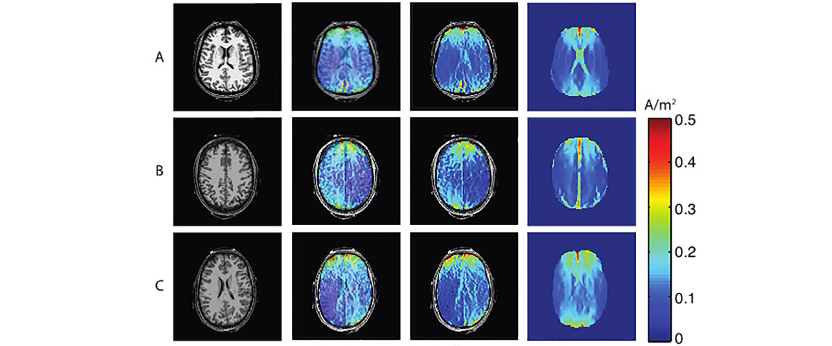
A MagLab user collaboration among three institutions used a novel application of magnetic resonance imaging (MRI) to measure the first images of the electrical current distribution within the living brain while the brain is experiencing transcranial electrical stimulation.
Why is this important?
Transcranial electrical stimulation has been shown to improve cognitive, motor and memory performance in healthy people. There is evidence that it may also provide new treatments for stroke rehabilitation and epilepsy. However, transcranial electrical stimulation is not well understood, in part because, before this study, no one could image the electrical current distribution in brains experiencing transcranial electrical stimulation. This new MRI method will be useful for improving the reproducibility and safety of transcranial electrical stimulation, as well as for advancing our ultimate understanding of the mechanisms that cause transcranial electrical stimulation to work.
Who did the research?
A. K. Kasinadhuni1, A. Indahlastari2, M. Chauhan2, M. Schar3, T. H. Mareci1, R. J. Sadleir2
1University of Florida; 2Arizona State University; 3Johns Hopkins University;
Why did they need the MagLab?
MRI research capabilities in the MagLab's AMRIS Facility developed this imaging method, which required the construction of control hardware interfaced to a 3T MRI system, as well as the development of data acquisition and data processing methodologies.
Details for scientists
- View or download the expert-level Science Highlight, Imaging current flow in the brain during transcranial electrical stimulation
- Read the full-length publication, Imaging of current flow in the human head during transcranial electrical therapy., in Brain Stimulation.
Funding
This research was funded by the following grants: G.S. Boebinger (NSF DMR-1157490); R. J. Sadleir (NIH R21 NS081646)
For more information, contact Joanna Long.


